Each year, 54,000 new cases of breast cancer are diagnosed in France. It is the most common type of cancer in women and also the most diagnosed cancer in the world. Nearly one in nine women will be affected by breast cancer in her lifetime. And the risk increases after age 50.
If a breast ultrasound/mammography performed for a routine exam or a suspicious lesion reveals an abnormality, it is followed up with a pathological analysis of the biopsied tissue (“path” report) and the patient waits for an accurate diagnosis. In the meantime, the patient embarks on a trying and stressful journey.
For the well-being of the patient and the effectiveness of surgery, how can the care pathway be accelerated and improved?
Breast cancer: what is the path report?
When breast cancer is suspected, the oncologist performs a biopsy (a surgical procedure under local anesthesia) which provides an accurate diagnosis and an appropriate treatment. The surgeon removes the suspicious tissue and sends it to the pathology lab to be examined under a microscope.
In the care pathway of a patient, the pathological analysis, abbreviated as the path report, is the only exam that conclusively confirms the diagnosis of breast cancer.
Breast cancer biopsy: what is the role of the sentinel lymph node?
Once breast cancer is diagnosed, the oncologist must assess the extent of the tumor to establish the most appropriate treatment plan for the patient. Thanks to additional imaging procedures, the surgeon can also know if the cancer cells have spread to other organs. Breast cancer treatment plans are the subject of a multidisciplinary collaboration also known as a multidisciplinary consultation meeting.
Removal of the sentinel lymph node is a procedure in which the first lymph node that drains the tumor is detected and dissected.
The path exam then determines whether these lymph nodes contain (or don’t contain) cancer cells, i.e., metastases (called micrometastases or macrometastases). Based on this exam, the cancer surgeon will decide whether or not further treatment is needed after surgery.
If the lymph node is “positive”, the surgeon can propose lymph node dissection which is often combined with drug therapy or radiotherapy.
Impact of sentinel lymph node biopsy techniques on the patient’s care pathway
Sentinel lymph node biopsy is the cornerstone of the care pathway. In this delicate procedure requiring the surgeon to use the best possible techniques to detect and dissect it.
Several techniques are used for sentinel lymph node detection:
- Isotope mapping: A radioactive marker is injected into the breast to identify the sentinel lymph node using a gamma probe. Radioactivity level is detected by sound.
- Colorimetric method (blue dye): Blue dye is injected into the breast and colors the lymphatics and sentinel lymph node which are visible to the naked eye.
- Combination method: this is the reference technique. It combines the isotope method (radioactive marker) with the colorimetric method (blue dye).
Nuclear medicine imaging, an effective but less convenient technique
In the isotope or combination method, a radioactive product is injected into the breast to enable detection and dissection of the lymph node.
To do this, the patient must go to a nuclear medicine center before her biopsy procedure.
Although conventionally used, this technique has some constraints and limitations:
- The solution must be injected before the biopsy procedure, usually the day before and no later than 90 minutes before the procedure.
- The appointment at the nuclear medicine center – which gives the injection – must therefore be coordinated with the biopsy schedule.
- Not all hospitals licensed to perform breast cancer surgery necessarily have a nuclear medicine department, which means that the patient may have to get her injection at another center, which may be far away.
- The patient is exposed to ionizing radiation (radioactivity).
- The medical team (nurses, doctors, surgeons) is also repeatedly exposed to the radioactivity present in the patients.
- The blue dye that is used can leave a tattoo mark on the skin, which may remain visible for several months or even years.
- The blue dye causes allergic reactions fairly frequently.
This complicated logistics is an additional source of stress for the patient and may result in delays in the care pathway.
Fluorescence imaging, the alternative for an accurate and inexpensive sentinel lymph node biopsy
To simplify the care pathway for patients with breast cancer, the National Cancer Institute recognizes the need for new, more efficient detection methods. The FLUOBEAM® fluorescence imaging system is one of these innovative techniques used in many hospitals and institutions including Hôpital Saint-Louis, and Hôpital Européen George Pompidou in Paris.
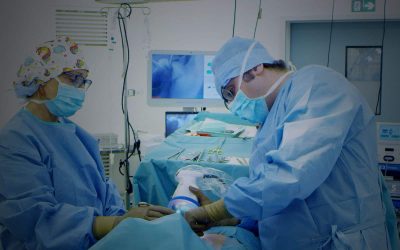
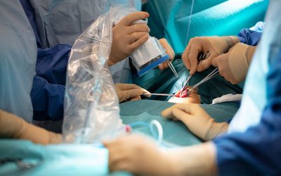
Thanks to injection of indocyanine green, this imaging technique allows the sentinel lymph node to be very clearly visualized by fluorescence. The system is designed to fit in the operating room environment to provide cancer surgeons with accurate, real-time images and information that is invisible to the naked eye: tissue perfusion, lymph node mapping, etc.
“Indocyanine green for detection of the sentinel lymph node in early breast cancer is feasible. It is accurate, safe and cheap”, explains Dr Charlotte Ngo Chirurgie, a Gynecological and Breast Cancer Surgeon at Hôpital Européen Georges Pompidou, Paris, France.
FLUOBEAM® alternative has several advantages:
- Performed in real time in the operating theatre and by the surgeon him/herself, it optimizes patient management:
- quick to set up,
- easy to use,
- immediate visualization allowing sentinel lymph node biopsy.
- No additional stress for the patient.
- No exposure to ionizing radiation.
Once breast cancer is diagnosed, sentinel lymph node biopsy is a fundamental step. Analysis of the biopsied tissue will determine the future treatment plan. Alongside the conventional methods for performing sentinel node biopsy, the fluorescence imaging system developed by FLUOPTICS(S) (FLUOBEAM®) is an effective alternative which optimizes the patient’s care pathway.
Additional reading to go further:
Breast reconstruction post-mastectomy: the alternative to breast implants.


