THYROID AND PARATHYROID SURGERY
Discover FLUOBEAM® LX METHODOLOGICAL APPROACH
AND OPERATING STRATEGY WITH FLUOBEAM® LX?
Autofluorescence imaging makes detection of parathyroid glands easier. Faster localization facilitates thyroid dissection and enables improved vascularization preservation. Parathyroid glands are usually identified after thyroid medialization, which reduces the risk of inadvertent resection.
Fluobeam® lx in action
Identification of parathyroid glands
Autofluorescence visualization of parathyroid adenomas, which guides and facilitates resection, is also enabled by FLUOBEAM® LX.
Identification of parathyroid glands
The natural fluorescence of parathyroid glands in the near infrared spectrum is called autofluorescence. FLUOBEAM® LX allows surgeons to identify these parathyroid glands in real time, which facilitates efforts to preserve the glands during the intervention.
Autofluorescence visualization of parathyroid adenomas, which guides and facilitates resection, is also enabled by FLUOBEAM® LX.
Identification of vessels vascularizing the parathyroid glands
Parathyroid glands and their vessels can be clearly identified by combining autofluorescence and perfusion visualization. This helps surgeons preserve the vessels during thyroid dissection.
The collective information, including localization and vascularization of the parathyroid glands, can make it possible to significantly reduce complications related to parathyroid lesions.
Checking the vascularity of the parathyroid glands
It is commonly accepted that complications during thyroid surgery, such as temporary hypocalcemia, are caused by inadvertent excision of the parathyroid glands or alteration of their vascularity.
Following intravenous injection of Indocyanine Green, FLUOBEAM® LX enables surgeons to check that the parathyroid glands are sufficiently perfused at the end of the procedure. This becomes valuable input for estimating their functionality.
TESTIMONIALS
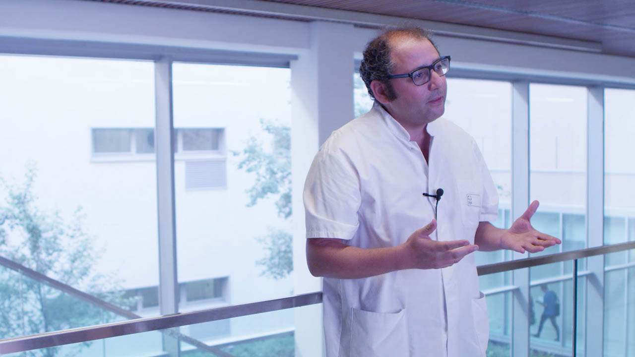
Dr. Fares BENMILOUD shares his experience with the FLUOBEAM® LX.
European Hospital, Marseille – France
Professor Frédéric TRIPONEZ shares his experience in fluorescence imaging
University Hospitals of Geneva – Switzerland
DISCOVER THE WEB MASTERCLASS
Don’t miss the opportunity to interact live with an expert on fluorescence in thyroid surgery. Register for a free one-hour session to discover or refine your knowledge of fluorescence imaging applied to thyroid surgery.
PUBLICATIONS
FLUOBEAM® LX is intended to provide real-time near infrared (NIR) fluorescence imaging of tissue during surgical procedures. The FLUOBEAM® LX is intended to assist in the imaging of parathyroid glands and can be used as an adjunctive method to assist in the location of parathyroid glands due to the auto-fluorescence of this tissue. The FLUOBEAM® LX is indicated for use in capturing and viewing fluorescent images for the visual assessment of blood flow in adults as an adjunctive method for the evaluation of tissue perfusion. This is a class II medical device. Product manufactured by FLUOPTICS SAS, France. For proper use, please read carefully all instructions in the specific instructions for use for each product.


